Brainstem Glioma Symptoms: MRI, DIPG, and DMG Explained
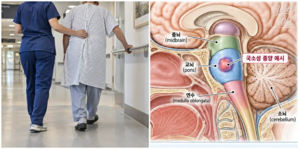
Brainstem glioma symptoms can look very different from one person to another because the brainstem contains pathways and cranial nerve structures involved in eye movement, facial movement, speech, swallowing, strength, balance, and other vital functions. Double vision, facial weakness, slurred speech, trouble swallowing, arm or leg weakness, and an unsteady gait can occur, but none of these symptoms by itself proves that a brainstem tumor is present.
The term brainstem glioma is also broader than many people realize. It describes gliomas that arise in the brainstem, while diffuse midline glioma, H3 K27-altered is a specific tumor diagnosis based on location plus molecular and pathologic features. DIPG, or diffuse intrinsic pontine glioma, is a clinical and imaging term for a diffuse tumor centered in the pons. These terms overlap, but they are not interchangeable.
Brainstem Glioma Symptoms Depend on Where the Tumor Is
The brainstem includes the midbrain, pons, and medulla. A tumor in one area may affect eye movements or facial muscles, while another may interfere more with swallowing, speech, limb strength, or coordination.
Common brainstem glioma symptoms can include:
- double vision or abnormal eye movements
- facial weakness or asymmetry
- slurred or changed speech
- difficulty chewing or swallowing
- weakness in an arm, leg, or one side of the body
- loss of balance, clumsiness, or trouble walking
- headache, nausea, vomiting, or unusual sleepiness if cerebrospinal fluid flow is blocked and hydrocephalus develops
Diffuse midline glioma can produce similar neurologic problems. The National Cancer Institute lists double vision, swallowing difficulty, weakness, and loss of balance among possible symptoms, with headache, vomiting, blurred or double vision, and sleepiness among symptoms that can occur when hydrocephalus raises pressure inside the skull.
The pattern matters more than one isolated complaint. A headache or a brief spell of dizziness is common and can have many causes. Brainstem glioma symptoms become more concerning when a specific neurologic change persists, progresses, or several related problems appear together.
Sudden Weakness or Slurred Speech Is an Emergency
A brain tumor is usually not the first condition to assume when facial weakness, arm or leg weakness, speech difficulty, vision changes, or severe imbalance begins suddenly. Stroke and transient ischemic attack can cause many of the same neurologic changes and require immediate assessment.
Sudden dizziness, weakness, or fainting can also have causes unrelated to a brain tumor, so it helps to understand how vasovagal syncope differs from orthostatic hypotension.
In the United States, call 911 for sudden one-sided weakness or numbness, new trouble speaking, sudden vision changes, sudden loss of balance or coordination, or a sudden severe headache. The CDC emphasizes that every minute counts during a stroke.
This distinction matters because brainstem glioma symptoms are often discussed as tumor warning signs, but the speed of onset changes the medical priority. New symptoms that appear over seconds or minutes should be treated as an emergency rather than watched at home to see whether they improve.
If symptoms are not sudden but double vision, facial asymmetry, swallowing difficulty, limb weakness, or gait changes continue or gradually worsen, a neurologic examination is still appropriate.
Why MRI Is the Main Imaging Test
When the neurologic examination suggests a possible brainstem lesion, brain MRI is the main imaging study. MRI with and without contrast can show where a lesion is centered, how much of the brainstem is involved, whether the growth pattern looks focal or diffuse, and whether nearby structures are affected.
For suspected pediatric glioma, the NCI describes MRI with and without contrast as part of the initial diagnostic evaluation. MRI is also central to the evaluation of DIPG and diffuse midline glioma because the location and growth pattern help narrow the diagnosis.
A CT scan may still be useful in an emergency, especially when clinicians need a rapid look for bleeding, major swelling, or hydrocephalus. But MRI provides the more detailed evaluation used to characterize a suspected brainstem tumor.
MRI can strongly suggest a diagnosis, but imaging alone cannot always establish the exact tumor type. In adults, the NCI notes that diffuse midline glioma cannot be diagnosed by imaging alone because other high-grade brain tumors can have a similar appearance.
Does Everyone Need a Biopsy?
Not always. The decision depends on the location of the lesion, its MRI appearance, the risks of the procedure, and whether tissue results would change treatment.
Historically, some tumors with classic DIPG imaging features were diagnosed without biopsy because of their location in the pons. Histologic confirmation is now obtained more often for molecular characterization and research or treatment planning, although the risks of a pontine biopsy still need to be considered carefully.
When tissue can be obtained safely, a neuropathologist can evaluate the tumor and molecular testing can look for changes such as H3 K27 alterations. This is especially important because the modern diagnosis of diffuse midline glioma, H3 K27-altered is not based on location alone. NCI describes this tumor entity as CNS WHO grade 4.
For an individual patient, the useful question is not simply, “Can this tumor be biopsied?” It is whether the expected diagnostic benefit is worth the procedural risk in that specific location and clinical situation.
Brainstem Glioma, Diffuse Midline Glioma, and DIPG Are Not the Same
These terms describe different aspects of a tumor.
| Term | What the term describes | Typical location or scope |
|---|---|---|
| Brainstem glioma | Broad location-based term for a glioma arising in the brainstem | Midbrain, pons, or medulla |
| Diffuse midline glioma, H3 K27-altered | Specific pathologic and molecular tumor entity | Midline structures such as the pons, thalamus, or spinal cord |
| DIPG | Clinical and imaging term for a diffuse infiltrating glioma centered in the pons | Pons |
A brainstem glioma can be focal or diffuse and can represent more than one tumor type. A diffuse midline glioma can arise in the brainstem, but it can also occur in other midline locations, including the thalamus or spinal cord. DIPG is centered in the pons and overlaps strongly with diffuse midline glioma, H3 K27-altered, particularly in children.
This is why brainstem glioma symptoms do not tell you which of these diagnoses is present. Symptoms show which neurologic functions are being affected. MRI shows the location and growth pattern. Tissue and molecular testing, when appropriate, provide the classification needed for a more precise diagnosis.
Why the Exact Diagnosis Matters for Treatment
Treatment cannot be decided from brainstem glioma symptoms alone. The tumor’s location, growth pattern, age of the patient, surgical accessibility, pathology, and molecular findings all affect the plan.
Some focal brainstem tumors can be considered for surgical removal or biopsy when their location allows it. Diffuse tumors in the pons are much harder to remove widely because tumor cells can intermingle with structures that control critical neurologic and bodily functions.
For diffuse midline glioma, radiation therapy remains an important part of treatment. The exact plan varies with age, tumor location, prior treatment, and available clinical trials.
There is also an important recent U.S. treatment update. On August 6, 2025, the FDA granted accelerated approval to dordaviprone (Modeyso) for adults and children age 1 year and older with diffuse midline glioma harboring an H3 K27M mutation whose disease has progressed after prior therapy. It was the first FDA-approved systemic therapy for H3 K27M-mutant diffuse midline glioma.
That approval does not mean Modeyso is approved for every brainstem glioma, every newly diagnosed patient, or every tumor called DIPG. The labeled indication requires the specified mutation and progressive disease following previous treatment.
That difference is another reason precise classification matters. A broad anatomic label may describe where the tumor is, while the molecular diagnosis can determine whether a particular treatment or clinical trial is relevant.
What to Bring to a Neurologic Evaluation
Because brainstem glioma symptoms can overlap with stroke, vestibular disorders, inflammatory disease, other tumors, and several neurologic conditions, a detailed timeline gives the clinician useful information when deciding what needs to be tested.
Before an appointment, write down:
- the first date and time each symptom appeared
- whether the change was sudden or gradual
- whether symptoms are constant, intermittent, or worsening
- which side of the face or body is affected
- whether double vision changes when one eye is covered
- whether swallowing problems occur with liquids, solids, or both
- whether there have been falls, new clumsiness, or trouble walking
- any headache, vomiting, unusual drowsiness, or change in alertness
- current medications and major medical history
This information cannot diagnose a brainstem glioma, but it gives the neurologic examination and imaging findings useful context.
The Bottom Line
Brainstem glioma symptoms reflect which structures in the brainstem are affected, so vision, facial movement, speech, swallowing, strength, and balance can all change. Persistent or progressive combinations of these problems deserve medical evaluation, while sudden weakness, speech difficulty, vision changes, or severe imbalance should be treated as a possible stroke emergency.
MRI is the main imaging test for a suspected brainstem tumor, but the final diagnosis may require pathology and molecular testing. Brainstem glioma is a broad location-based term, diffuse midline glioma is a specific molecular-pathologic diagnosis, and DIPG describes a diffuse tumor centered in the pons. They can overlap, but they are not the same diagnosis.
Source:
- National Cancer Institute — Diffuse Midline Glioma: Diagnosis and Treatment
- National Cancer Institute — Childhood Astrocytomas and Other Gliomas Treatment (PDQ)
- National Cancer Institute — Diffuse Intrinsic Pontine Glioma (DIPG)
- Centers for Disease Control and Prevention — Signs and Symptoms of Stroke
- U.S. Food and Drug Administration — FDA Grants Accelerated Approval to Dordaviprone for Diffuse Midline Glioma