Pyoderma in Dogs: Symptoms, Scabs, Causes, Treatment
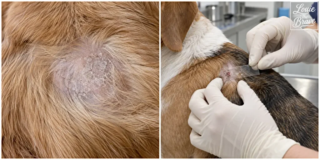
Pyoderma in dogs is a bacterial skin infection that can cause scabs, scaling, red bumps, pustules, circular areas of peeling skin, and patchy hair loss. Those signs can look similar to allergies, ringworm, mites, or other skin diseases, so appearance alone cannot confirm the diagnosis.
The most common form is superficial Pyoderma in Dogs, also called superficial bacterial folliculitis. It affects the epidermis and the upper part of the hair follicle. Deep pyoderma extends farther into the skin and is more likely to cause pain, swelling, odor, draining pus or blood, ulcers, and thick or bloody crusts. Merck notes that Staphylococcus pseudintermedius is the primary bacterial pathogen associated with canine pyoderma.
What Does Pyoderma in Dogs Look Like?
Superficial infection often starts around hair follicles. A dog may develop small red papules, pustules, crusts, scales, or patchy areas of hair loss. One especially useful clue is an epidermal collarette: a round or ring-shaped area where the center has cleared and the edge has a rim of peeling scale or crust.
These lesions are often found on the trunk or underside of the body, but they can appear elsewhere. Itching varies. Some dogs scratch or lick frequently, while others have visible lesions without intense itching.
| What you may see | What it can mean |
|---|---|
| Flakes or scaling around hairs | Can occur with superficial pyoderma, but also with other skin disorders |
| Red bumps or pustules | May reflect bacterial folliculitis, but are not diagnostic by themselves |
| Circular crusts or peeling rings | May be epidermal collarettes seen with superficial pyoderma |
| Patchy hair loss | Can occur when inflamed follicles lose hair |
| Pain, odor, pus, blood, or draining tracts | More concerning for deep infection |
A photo can help document how a lesion changes, but it cannot show whether bacteria are actually causing the inflammation.
Why Scabs and Flakes Do Not Prove It Is Pyoderma in Dogs
Scabs, scaling, redness, and hair loss are not unique to bacterial infection. Dermatophytosis, commonly called ringworm, and demodicosis caused by Demodex mites can produce overlapping changes. Allergic skin disease can also cause a dog to scratch and lick until the skin barrier is damaged, making a secondary bacterial infection more likely.
This is why veterinarians often use skin cytology. A sample from the lesion is examined under a microscope for bacteria and inflammatory cells. Depending on the pattern, a veterinarian may also perform skin scrapings, fungal testing, or other dermatologic tests to rule out mites, dermatophytes, yeast, and other causes.
If the infection keeps returning, the underlying problem matters as much as the infection itself. Recurrent nonseasonal itching, paw licking, ear inflammation, or repeated bacterial and yeast infections can be part of an allergy workup. A controlled elimination diet may be discussed when food allergy is suspected; the process is explained in this guide to hydrolyzed dog food and elimination diets.
What Causes Pyoderma in Dogs?
Healthy canine skin already carries bacteria. Pyoderma usually develops when something changes the skin environment or damages the protective barrier enough for bacteria to overgrow and invade.
Common predisposing problems include allergic dermatitis, fleas and other ectoparasites, demodicosis, repeated scratching or licking, poor grooming, keratinization disorders, and warm, moist skin folds. Endocrine diseases such as hypothyroidism and hyperadrenocorticism, also called Cushing’s disease, are especially relevant when infections are recurrent or difficult to clear.
Moisture can also matter. Folds around the lips, face, neck, armpits, feet, vulva, and tail can stay warmer and more humid than exposed skin. Merck notes that these areas often have higher bacterial counts and may be more prone to infection.
If recurrent skin infections occur together with increased thirst and urination, increased appetite, panting, a pot-bellied appearance, muscle loss, or thin skin, a veterinarian may consider endocrine testing. The diagnostic process for hyperadrenocorticism is covered in this guide to Cushing’s disease testing and monitoring in dogs.
How Vets Diagnose Pyoderma
The goal is not simply to decide that a lesion “looks bacterial.” Diagnosis is stronger when the lesion pattern fits and cytology shows bacteria together with inflammatory cells.
For a first uncomplicated case, cytology may be enough to support treatment. When pyoderma in dogs keeps returning, the diagnostic plan usually has to go beyond the visible scabs and bumps. Bacterial culture and susceptibility testing become more important when infection is deep, repeatedly returns, has failed previous treatment, or follows repeated antimicrobial exposure.
The 2025 WSAVA and ISCAID guidance specifically emphasizes cytology, selective culture, topical therapy, and finding the primary cause rather than automatically reaching for systemic antibiotics.
Culture can identify which bacteria are present and which antibiotics are likely to work. That matters because methicillin-resistant Staphylococcus pseudintermedius, or MRSP, and other resistant bacteria can make treatment more complicated.
Does Every Dog Need Oral Antibiotics?
No. Current antimicrobial guidance recommends topical antimicrobial therapy as the treatment of choice for surface and superficial pyoderma when it can be applied effectively. Systemic antibiotics are generally reserved for deeper infections, cases that do not respond adequately to topical treatment, or situations in which topical therapy is not practical.
Topical treatment may include veterinary shampoos, mousses, sprays, wipes, or other products. Chlorhexidine is one commonly used active ingredient, and clinical studies cited in the 2025 ISCAID guideline support several 2% to 4% chlorhexidine formulations for superficial bacterial folliculitis. The right concentration and contact method depend on the product and the dog’s skin condition.
Do not substitute a human disinfectant or improvise a dilution at home. Use a veterinary product recommended for the dog’s condition and follow the application directions. Stop and contact the veterinary clinic if a topical product causes marked irritation.
Should You Pick Off the Scabs?
Avoid pulling off a crust simply to see what is underneath. A firmly attached scab can cover inflamed or damaged skin, and forcing it off may cause bleeding, pain, or additional trauma.
More than 25 years ago, I found a hard crust on my dog Ppoto’s back and pulled it off without thinking much about it. The skin underneath looked much deeper and more damaged than I expected. A veterinarian gave us an ointment and the area eventually healed, but the lesion was never specifically diagnosed as pyoderma. That old experience is a good reminder that a scab’s appearance does not tell you what is happening below the surface.
The same caution applies to leftover medication. An old antibiotic or prescription ointment may have been intended for a different problem. Reusing it on a new lesion can delay the correct diagnosis and may contribute to inappropriate antimicrobial exposure.
If a dog keeps licking or scratching the area, an Elizabethan collar or recovery garment may help prevent additional self-trauma while the cause is being addressed. It protects the skin; it does not treat the infection itself.
When Does Pyoderma Need a Vet Visit?
A small flaky patch is not automatically an emergency, but veterinary examination becomes more important when the lesion is painful, rapidly expanding, repeatedly returning, producing pus or blood, developing odor, or causing significant hair loss or itching. Deep pyoderma can produce swelling, ulcers, hemorrhagic crusts, draining tracts, and purulent or bloody discharge.
Prompt evaluation is also reasonable when several lesions appear at once, the dog seems unwell, or home care has not stopped the area from worsening. Recurrent pyoderma deserves a search for the trigger instead of repeated treatment of the visible infection alone.
After medicated bathing, dry the coat and skin thoroughly so damp areas do not stay wet for long periods. For dogs with skin folds, paws that remain moist, or dense coats, moisture control can be part of preventing repeated bacterial overgrowth.
Is Pyoderma Contagious?
Most canine pyoderma is not considered a contagious disease passed from one dog to another. The bacteria most often involved, especially S. pseudintermedius, commonly live on healthy dogs and usually become a problem when the skin barrier or underlying health changes.
However, bacterial strains can occasionally be shared between animals and people, and resistant strains deserve extra care. Good hand hygiene and avoiding direct contact with draining material are sensible, especially for immunocompromised household members.
Bottom Line
Pyoderma in dogs can look like scabs, flakes, red bumps, circular crusts, or patchy hair loss, but those signs are not specific enough to diagnose a bacterial infection at home. Cytology helps confirm whether bacteria and inflammation are present, and recurrent cases may need culture plus testing for allergies, parasites, or endocrine disease.
For superficial infections, topical antimicrobial treatment is now preferred whenever it is appropriate and practical. Deep, painful, draining, recurrent, or treatment-resistant lesions need a veterinary plan that addresses both the infection and the reason the skin keeps becoming vulnerable.
Source: